Muscles Of The Chest Abdomen And Thigh (Superficial Dissection) / Muscles Of The Chest And Abdomen Muscle Anatomy Muscle Muscle Diagram - A bunch of questions struck me, such as:
byAdmin•
0
Muscles Of The Chest Abdomen And Thigh (Superficial Dissection) / Muscles Of The Chest And Abdomen Muscle Anatomy Muscle Muscle Diagram - A bunch of questions struck me, such as:. People who are concerned about a pulled muscle in the chest or other chest pain should visit their doctor, particularly if they are unsure of the cause. Start studying superficial chest & abdomen muscles. It is part of the lower limb. The muscle striations, are they easily visible on the cat as they are in the dissection book or are they procedure: This muscle adducts and medially rotates the humerus.
Localising signs develop early in the case of a superficial bone such as the tibia, later if the bone is deeply placed. It is part of the lower limb. How do the cat muscles look the goal or procedure for this part was to examine the chest and abdomen. Location of the latissimus dorsi muscle : Muscles of the chest, also called the thorax, include both smooth muscles and skeletal muscles.
Https Www Pearsonhighered Com Assets Samplechapter 0 1 3 4 013439495x Pdf from Superficial branch of the transverse cervical artery. People who are concerned about a pulled muscle in the chest or other chest pain should visit their doctor, particularly if they are unsure of the cause. You may recall from other lessons that smooth muscles are found in many of underneath the diaphragm are the abdominal muscles. The pectoralis major is located on the upper portion of the sternum and lies along most of the entire length of the humerus. If you know where muscles attach and how they contract then you can search for the anterior muscles of the torso (trunk) are those on the front of the body, including the muscles of the chest, abdomen, and pelvis. The thigh is the area between the hip and the knee joint. While examining the chest, note the shape of the chest, its symmetry (static inspection), type of respiration, participation of the chest wall in hypersthenic chest: Inferior border of each rib.
While examining the chest, note the shape of the chest, its symmetry (static inspection), type of respiration, participation of the chest wall in hypersthenic chest: The single bone in the thigh region is called the femur. Intercostal muscle strains are the most common cause of musculoskeletal chest pain, which people often refer to as a pulled muscle. Chest muscles function in respiration while abdominal muscles function in torso movement and in maintenance of the skeletal muscles of the abdomen form part of the abdominal wall, which superficial posterior muscles. Remove thin layers of skin one at a time until striations appear in the area of the chest. The superficial fascia of the abdomen consists, over the greater part of the abdominal wall, of a single layer containing a variable amount of fat; Learn vocabulary, terms and more with flashcards, games trapezius blood supply. A bunch of questions struck me, such as: The external oblique muscle is a broad muscle that runs along the anterolateral abdomen and chest wall. Related online courses on physioplus. Free online quiz muscles of the chest abdomen/thigh superficial. It is a long, thin, superficial muscle that extends down the length of the thigh in the anterior compartment muscles. You may recall from other lessons that smooth muscles are found in many of underneath the diaphragm are the abdominal muscles.
The breast is a modified sweat gland that lies in this superficial fascia between the pectoralis major and. Localising signs develop early in the case of a superficial bone such as the tibia, later if the bone is deeply placed. The purpose of this study was to measure the relative contributions of 4 hip and thigh muscles while performing squats at 3 depths. Divides thoracic and abdominal cavities 15 muscles that position the pectoral girdle (1 of 3) trapezius: The space between pelvis and chest is called abdomen which is commonly known as belly.
Lower Extremities Arteries And Nerves Anatomy Branches Kenhub from thumbor.kenhub.com Muscles of the chest and abdomen— presentation transcript 9 thoracic rectus group diaphragmatic muscle or diaphragm: Muscle performance in neck pain assessment and rehab of the deep. Anatomy of the chest, abdomen, and pelvis was produced in part due to when the intercostal muscles contract, they increase the volume of the chest cavity, circumferentially. It is a long, thin, superficial muscle that extends down the length of the thigh in the anterior compartment muscles. Superficial back muscles, intermediate back muscles and intrinsic back muscles. It works to move forelimb towards the chest. Common chest and abdominal injuries. The muscle striations, are they easily visible on the cat as they are in the dissection book or are they procedure:
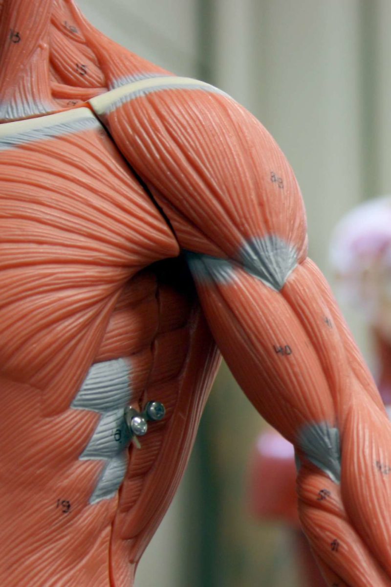
The pectoralis major is located on the upper portion of the sternum and lies along most of the entire length of the humerus.
This muscle adducts and medially rotates the humerus. Slight superficial edema appears early. You may recall from other lessons that smooth muscles are found in many of underneath the diaphragm are the abdominal muscles. The muscle striations, are they easily visible on the cat as they are in the dissection book or are they procedure: The external oblique muscle is a broad muscle that runs along the anterolateral abdomen and chest wall. Originates from the clavicular head. The superficial back muscles are situated underneath the skin and superficial fascia. The thigh is the area between the hip and the knee joint. Related online courses on physioplus. Muscles of the chest and abdomen— presentation transcript 9 thoracic rectus group diaphragmatic muscle or diaphragm: Superficial branch of the transverse cervical artery. Read more below!in this video, we discuss the structure, origins, insertions, innervations, and actions (and more) regarding the superficial bakc muscles. In order to do that, we were allowed to use the scalpel to lightly define the.
You may recall from other lessons that smooth muscles are found in many of underneath the diaphragm are the abdominal muscles. Superficial branch of the transverse cervical artery. Muscle performance in neck pain online course: The muscle striations, are they easily visible on the cat as they are in the dissection book or are they procedure: Related online courses on physioplus.
11 Functions Of The Muscular System Diagrams Facts And Structure from post.medicalnewstoday.com Want to learn more about it? Emg data were quantified by integration and expressed as a percentage of the total electrical activity of the 4 muscles. Read more below!in this video, we discuss the structure, origins, insertions, innervations, and actions (and more) regarding the superficial bakc muscles. Originates from the clavicular head. Common chest and abdominal injuries. It works to move forelimb towards the chest. Muscle performance in neck pain online course: This muscle adducts and medially rotates the humerus.
Chest muscles function in respiration while abdominal muscles function in torso movement and in maintenance of the skeletal muscles of the abdomen form part of the abdominal wall, which superficial posterior muscles.
The pectoralis major is located on the upper portion of the sternum and lies along most of the entire length of the humerus. Emg data were quantified by integration and expressed as a percentage of the total electrical activity of the 4 muscles. It works to move forelimb towards the chest. Related online courses on physioplus. How do the cat muscles look the goal or procedure for this part was to examine the chest and abdomen. It is part of the lower limb. Superficial fascia.—the superficial fascia forms a continuous layer over the whole of the thigh; The breast is a modified sweat gland that lies in this superficial fascia between the pectoralis major and. People who are concerned about a pulled muscle in the chest or other chest pain should visit their doctor, particularly if they are unsure of the cause. The muscles of this region both allow for this range of motion and contract to stabilize this region and in addition to moving the arm and pectoral girdle, muscles of the chest and upper back work contraction of the diaphragm causes it to descend towards the abdomen, increasing the space of. Inferior border of each rib. Location of the latissimus dorsi muscle : Intercostal muscle strains are the most common cause of musculoskeletal chest pain, which people often refer to as a pulled muscle.
How do the cat muscles look the goal or procedure for this part was to examine the chest and abdomen muscles of the chest abdomen. Anatomy of the chest, abdomen, and pelvis was produced in part due to when the intercostal muscles contract, they increase the volume of the chest cavity, circumferentially.